
What is Benign Bone Lesion Treatment?
Benign bone lesion treatment is medical care rendered to a patient with a diagnosis of benign (noncancerous) bone lesion.
Bone lesions are defined as areas of bone that are damaged or changed as a result of tumors, infections, or fractures. Most bone lesions are benign or noncancerous in nature, usually non-life-threatening, and do not spread to other parts of the body. However, some bone lesions are malignant or cancerous in nature. These bone lesions are life-threatening and cause cancer cells to spread throughout the body. Bone lesions can occur in any part of the bone - from the surface to the marrow in the center - and can affect any area of the body.
Types of Benign Bone Lesions
Some of the types of benign bone lesions include:
- Aneurysmal bone cyst (ABC): Aneurysmal bone cyst is a highly destructive non-cancerous bone tumor of the blood vessels that originates in the bone marrow. It occurs as a blood-filled fibrous tumor-like cyst that enlarges the bone, giving it an inflated appearance. These cysts are rare and most often occur in patients from birth to twenty years of age. They most commonly develop in the leg, arm, spine, and pelvis.
- Unicameral (simple) bone cyst (UBC): Unicameral bone cyst is a simple solitary bone cyst found inside a bone as a cavity filled with straw-colored fluid. These cysts are typically seen in children younger than twenty years of age and commonly occur in the upper thighbone (proximal femur) or upper arm (proximal humerus). Less common sites of occurrence include the ankle, heel, or pelvis. Most cases of UBCs have no symptoms and are discovered accidentally during imaging tests.
- Nonossifying fibromas (NOFs): Nonossifying fibromas are the most common noncancerous bone tumor occurring in children under the age of 20. Boys are twice more likely to be affected with this tumor than girls. These tumors are mainly made up of scar (fibrous) tissue and are non-aggressive. NOFs most often occur in the thigh bone (femur) or the shin bone (tibia), but may also be found in the upper arm bone (humerus). Most cases of NOFs do not require any treatment as they resolve on their own when children fully develop.
- Fibrous dysplasia: Fibrous dysplasia is a congenital (present at birth) bone condition that affects bone growth and development. It is a rare bone disorder that causes formation of too much fibrous-type tissue in place of normal bone tissue due to the failure of bone-forming cells to mature. The fibrous tissue can weaken the affected bone and cause it to fracture or deform. The condition usually occurs in children in the age group of 3 to 15 and most commonly affects the upper arm, thigh bone, and shinbone. However, any bone in the body can be affected, including the pelvis, ribs, face, and skull.
- Osteochondroma: This is the most common type of benign bone lesion and affects the growing ends of long bones, such as the leg or arm bones. It is most often found in adolescents and teenagers. Osteochondromas account for 35 to 40 percent of all benign bone lesions. They are made up of cartilage and bone and are considered a growth abnormality. Children may develop a single osteochondroma or several of them.
Treatment for Benign Bone Lesions
Treatment for benign bone lesions depends on the type of tumor, its location and size, and the age of the patient.
In general, benign bone lesions only require close observation to see if the lesion changes over time. During this period, you may require regular follow-up x-rays or other tests to assess the status of the lesion. Benign bone lesions may not warrant any treatment unless it starts to interfere with your movement and everyday routine.
Some benign lesions can be effectively treated with the use of medications, although they can return.
Bone lesions can gradually grow, remain the same, or eventually disappear. Children have a high chance of having their bone lesions disappear as they develop.
However, in some cases, your physician may opt to surgically remove the lesion to prevent it from becoming malignant and spreading to other parts of the body.
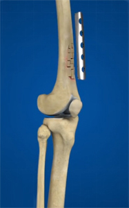
Benign bone tumors are commonly treated by a procedure called curettage. This involves scraping the lesion with an instrument called a curette. Following the procedure, blood fills the defect (hematoma) and bone matrix is produced in the hematoma. The curettage process activates the bone matrix which helps regenerate bone in the defect. Large bone defects may be reinforced with bone substitutes such as bone cement, bone grafts obtained from your body or an implant. Curettage also helps the bone integrate and fuse with a graft or implant.
For further destruction of tumor cells, along with curettage, adjuvant treatments such as liquid nitrogen, phenol, polymethylmethacrylate (PMMA), or thermal cautery may be administered.
Your physician may recommend surgical excision of a lesion if there is a risk that it may cause a fracture or disability. For tumors that are very large and may involve a joint, complete excision with joint reconstruction may be necessary.