
What is Distal Femoral Resection and Reconstruction?
Benign or malignant tumors can develop in or spread to the lower (distal) part of the femur (thighbone), causing progressive bone tissue destruction associated with pain and disability. Distal femoral resection is a procedure to remove the diseased distal femur, which can then be reconstructed with an artificial prosthesis to restore function. Such conditions were initially treated with above-knee amputations of the limb, but with modern medicine improving survival rates and advances in imaging, surgical technique, and implant design, limb salvage surgery now promises better functional and cosmetic outcomes.
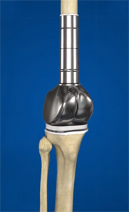
Modern prostheses for distal femoral reconstruction are modular, with components of different shapes and sizes. They allow flexion and extension as well as rotatory movements of the knee. Some prosthetic implants can even expand, which is necessary for patients who are skeletally immature. Custom-made implants may be recommended in cases with unusual anatomy.
Diagnosis
To diagnose your condition, your symptoms and medical history are reviewed, and a history and physical examination is performed. Diagnostic tests may include X-rays, bone scans, MRI, CT scans, and angiography to evaluate the neighboring popliteal blood vessels. In certain cases, a biopsy of a lesion may be needed to make a clear diagnosis. Cancer staging is assessed in the case of malignant tumors. If the tumor is found to be a metastatic lesion, tests are performed to locate the primary tumor.
Indications
Distal femoral resection and reconstruction is performed to completely remove the tumor in the distal femur. Your doctor will recommend this procedure after considering a number of factors, including the extent of the tumor, its location, radiographic features, presence of a fracture, the involvement of neurovascular structures, the underlying diagnosis, and expected survival.
Amputation may be considered if the tumor is considered unresectable due to infection or extensive tissue destruction
Preparation for the Procedure
Chemotherapy is usually performed prior to surgery to shrink the tumor and control its margins. Diagnostic studies help determine the extent of bone and soft tissue resection necessary, the proximity of the tumor to important nerves and vessels, and the dimensions of the prosthesis required.
Procedure for Distal Femoral Resection and Reconstruction
The procedure is carried out under general anesthesia, lying on your back (supine). A long incision is made from the middle of the inside thigh and extending down across the knee.
The lower hamstrings at the back of the thigh are exposed, detached, and retracted to locate the popliteal vessels and the sciatic nerve which are then protected during the surgery.
The distal femur is approached by creating a space between the overlying inner thigh muscles. The joint capsule of the knee is opened, and knee ligaments and menisci are removed. Resection of the distal femur is then performed as planned preoperatively with imaging studies. A wider margin of bone is removed in cases of sarcomas as compared to metastatic carcinomas. Your surgeon then measures the bone defect and chooses the appropriate prosthetic components.
An assembled modular type of prosthesis with femoral and tibial components is usually used for reconstruction. The medullary or central cavity of the femur is widened to allow insertion of the femoral component. Part of the tibial bone of the shin is also removed to allow insertion of the tibial component.
Postoperative Treatment
Following surgery, your leg will be elevated for about 3 days to prevent swelling. The wound is continuously suctioned through drainage tubes for 3-5 days during which time you will receive intravenous antibiotics. You will wear a brace to restrict the movement of the knee for 2-3 weeks to allow the skin and muscles to heal. Certain isometric exercises will be recommended during this time and you will usually be allowed to weight-bear early.
Risks and Complications
As with any surgery, distal femoral resection and reconstruction may be associated with certain risks and complications such as:
- Wound infection
- Implant loosening
- Implant breakage or failure
- Recurrence of the tumor
In most cases, distal femoral resection and reconstruction has good functional outcomes with good stability, and recurrence of the tumor at the distal femur is rare.