
What are Fractures in Cancer Patients?
A fracture is a painful condition where there is a break in one or more bones. Certain types of cancer have been connected with an increased risk of fractures. A high-risk group regarding fractures includes cancers that primarily affect the bones such as primary bone cancer, lung cancer, multiple myeloma, prostate cancer, cancer of the liver, gall bladder, and pancreas, breast cancer, metastases to the bone, and metastases to organs other than bone.
A few types of cancers that have been extensively studied for fracture risk include prostate cancer in men and breast cancer in women. Research on prostate cancer has shown that the use of anti-androgen therapy for prostate cancer can result in decreased bone mineral density and increased risk of fractures. Women with breast cancer have been noted with an increased risk of vertebral fractures.
Types of Fractures in Cancer Patients
Some of the types of fractures in cancer patients include:
- Transverse fracture: This type of fracture occurs along the bone shaft in a horizontal line pattern.
- Oblique fracture: In this type, the fracture occurs across the bone shaft in an angled line pattern.
- Spiral fracture: In this type, the fracture line surrounds the bone shaft and occurs as a result of a twisting force.
- Comminuted fracture: This type of fracture involves a splinter or break of bone into 3 or more pieces.
- Open fracture: This is also known as compound fracture and causes serious damage to the surrounding soft tissue structures as the bone fragments stick out through the skin to the external air exposing the fracture site.
Causes of Fractures in Cancer Patients
Bone is a form of connective tissue made of mainly minerals, such as calcium, and a form of protein known as collagen. Bone is always active and constantly repairs and renews itself with a process known as remodeling. Two types of bone cells assist with this process:
- Osteoblasts, cells that form or build new bone
- Osteoclasts, cells that reabsorb or break down old bone
When both these cells are functioning in the way they should, new bone is constantly formed while old bone is broken down. This helps to keep the bones strong.
However, when cancer cells speed up or block the action of the osteoclasts and osteoblasts, too much bone is formed or too much bone is broken down. Either of these changes can lead to weakening of the bones making them more prone to fracture and/or a dangerous escalation of calcium level in the blood (hypercalcemia).
Signs and Symptoms of Fractures in Cancer Patients
Some of the signs and symptoms of fractures in cancer patients include:
- Pain
- Swelling
- Bone loss
- Hypercalcemia
- Joint instability
- Joint deformity
- Impaired mobility
Diagnosis of Fractures in Cancer Patients
Diagnosis of fractures in cancer patients is made through evaluation of medical history, a physical examination, and other diagnostic imaging tests such as:
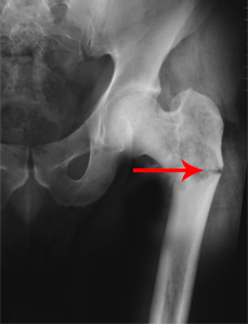
- X-rays: This study uses high electromagnetic energy beams to produce images of the bones and helps to detect whether the bone is intact or broken, and the type of fracture and its location.
- CT scan: This scan uses special x-rays that produce clear images of any damage present in the internal hard and soft tissue structures of the body that is not visible in an x-ray. It provides your doctor with crucial information about the severity of the fracture.
- Bone scan: This is a nuclear imaging study that helps your doctor to detect hidden stress fractures or any bone disorders such as avascular necrosis, arthritis, Paget’s disease of the bone, or bone metastasis. The scan examines the entire skeleton in your body, so all the bones can be checked for cancer.
- MRI Scan: This study utilizes large magnetic fields and radio waves to produce cross-sectional images of the bones and tissues. It is very beneficial in closely examining the spine, spinal cord, and joints and often provides a better view of the outline of a bone mass viewed on an x-ray.
Treatment of Fractures in Cancer Patients
Treatment for fractures in cancer patients depends upon the severity of the fracture and involves both non-surgical and surgical methods. These include:
Non-surgical methods
- Bisphosphonate Drugs: Use of bisphosphonate drugs can successfully prevent pathological fractures, alleviate bone pain from metastases, and reduce bone loss in individuals at risk of bone complications from cancer and its treatment. Bisphosphonate drugs function by preventing bone breakdown or resorption. Zometa® (zoledronic acid) and Aredia® (pamidronate) are the two most commonly employed bisphosphonate drugs for the treatment of cancer-related skeletal complications.
- Splint/Casting: This method is mostly employed as an initial treatment measure in which a splint may be used to provide support and comfort for the fractured bone. Your doctor may also recommend immobilizing your limb in a rigid cast for several weeks. The cast is then replaced with a functional brace which allows some movement of the limb while still providing support and protection until complete healing.
Surgical methods
- External fixation: This method is performed in case you have multiple injuries. Your doctor will place metal screws and pins inside the bone which will be attached to a rod present outside the skin. This helps in stabilizing the bones.
- Intramedullary nailing: In this method, a rod is passed along the broken bone and intramedullary nails made of titanium are fixed above and below the bone shaft to stabilize and heal it.
- Plates and screws: In this procedure, your doctor will reposition the fragmented bones and align them by fixing it with screws and metal plates. The plates and screws method is employed when intramedullary nailing is not a viable option.
Recovery post-surgery may take anywhere from 4 to 6 months or longer and varies from individual to individual depending on the severity of the fracture.